
The Significance of PET/CT in Neurology—Why It Matters Now
Positron Emission Tomography combined with low-dose CT (PET/CT) has moved from research labs into everyday neurologic care. It doesn’t just show the brain—it measures how it’s working. For patients and families, that means clearer answers, earlier, and care plans that are tailored to the biology of their condition.
Dr. Sherif Makar
11/2/20252 min read
Where PET/CT helps most in neurology
1) Memory loss & dementia syndromes
Two big advances make PET/CT especially relevant today:
Amyloid and tau PET: Amyloid PET detects plaque pathology; tau PET visualizes neurofibrillary tangles—together they anchor the modern “AT(N)” biomarker framework for Alzheimer’s disease (AD). Updated Appropriate Use Criteria (AUC) from the Society of Nuclear Medicine & Molecular Imaging (SNMMI) and the Alzheimer’s Association explain when these scans are most helpful (for example, unexplained MCI where results would change management).
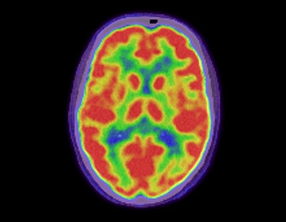
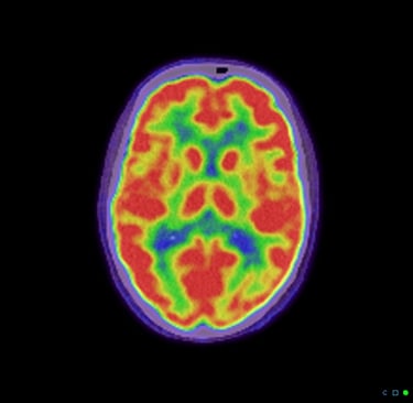
FDG PET metabolism: FDG (glucose) PET reveals characteristic patterns that help tell AD from frontotemporal dementia or dementia with Lewy bodies and can support diagnoses when MRI and labs aren’t definitive. New joint SNMMI/EANM guidelines detail these patterns and when to use FDG PET alongside amyloid/tau or CSF biomarkers.
Why it matters now: disease-modifying therapies for early AD (lecanemab; donanemab) require confirmed amyloid pathology and thoughtful risk–benefit discussion. PET/CT is one of the clearest paths to that confirmation.
2) Parkinsonism & atypical parkinsonian syndromes
F-18 FDOPA PET measures dopaminergic terminals in the striatum. In suspected parkinsonian syndromes, reduced putaminal uptake supports a neurodegenerative parkinsonism diagnosis. The FDA-approved label includes a simple carbidopa pre-med (usually 150 mg by mouth 60–120 minutes before the scan) and a standardized acquisition window, which helps produce consistent, high-quality results.
3) Epilepsy (especially when MRI looks “normal”)
FDG PET can localize an epileptogenic focus in focal epilepsy—information that is crucial for surgical candidacy and planning, particularly when structural imaging is negative. European and international guidance outlines how FDG PET fits into presurgical work-ups and monitoring.
4) Neuro-oncology & inflammatory disorders
While MRI remains first-line, PET/CT can help distinguish tumor recurrence vs. treatment effect and, with FDG, can sometimes flag neuroinflammation when clinical suspicion remains high. (Use is individualized and guided by specialist review.)
Coverage is evolving—in a good way
In October–November 2023, Medicare retired the national “coverage with evidence development” limit for beta-amyloid PET. Coverage decisions now rest with regional MACs, which has expanded access across much of the U.S. (Medicare Advantage plans set their own authorization rules). Practically, this means more eligible patients can receive amyloid PET as part of real-world care, not just trials. We verify benefits for each patient.
Safety & preparation
PET/CT uses a small amount of radioactivity—similar to other diagnostic imaging—and the CT portion is typically low-dose for brain scans. Most patients go home right after. Preparation varies by tracer: for FDOPA, we often use carbidopa pre-medication to optimize signal and image quality; for FDG, quiet rest and controlled blood glucose improve accuracy. We’ll give you clear, written instructions before your appointment.
What this means for patients at Charis Neurology
Faster, clearer answers when symptoms are subtle or diagnoses overlap.
Biology-based decisions about new Alzheimer’s therapies (which require proof of amyloid) and about management of parkinsonian syndromes.
Team-based reading: our neurologists integrate PET/CT with MRI, neuropsychological testing, labs, and your story—because imaging is most powerful when interpreted in context.
When do we recommend PET/CT?
We consider PET/CT when results are likely to change what we do next—for example:
Mild cognitive impairment where confirming or excluding AD pathology will guide treatment and planning.
Early parkinsonism with diagnostic uncertainty where dopaminergic imaging clarifies the picture.
Refractory focal epilepsy being evaluated for surgery.
The bottom line
PET/CT has become a practical, patient-centered tool in modern neurology. It helps us diagnose earlier, tailor treatments (including newly approved disease-modifying options for early Alzheimer’s), and make more confident decisions with you. If you or a loved one are navigating memory concerns, parkinsonian symptoms, or complex seizure disorders, talk with our team—together we’ll decide whether PET/CT is the right next step.

© 2025 Charis Neurology. All rights reserved.